Iliacus Muscle: Anatomical Details
The iliacus muscle is a flat, triangular muscle that is part of the iliopsoas group. It plays a vital role in hip flexion and stability, working in conjunction with the psoas major muscle. Understanding its anatomy can help in identifying its functions and addressing any related issues.
Anatomy of the Iliacus Muscle
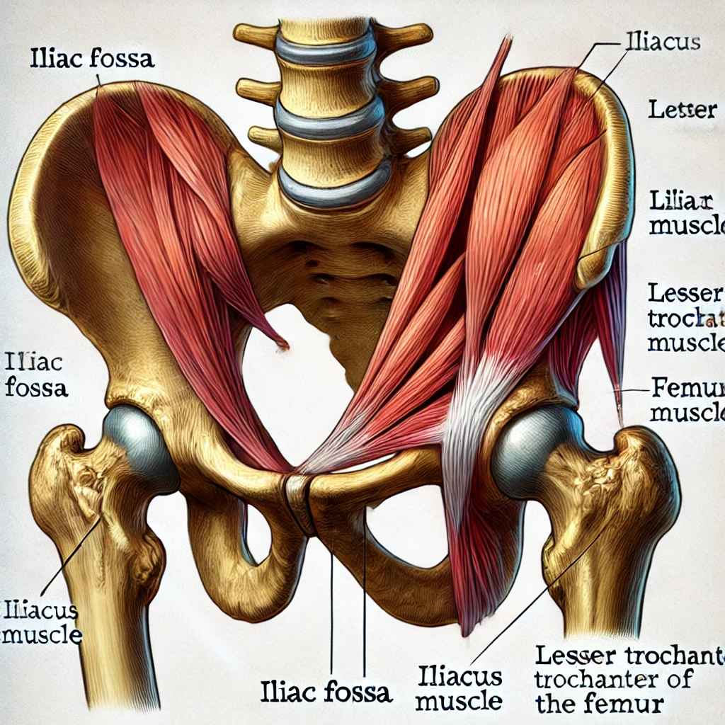
The iliacus muscle originates from the iliac fossa on the interior side of the hip bone and the ala of the sacrum. It inserts onto the lesser trochanter of the femur, combining with the tendon of the psoas major muscle. The iliacus is innervated by the femoral nerve (L2-L4) and receives blood supply from the iliolumbar artery.
Function
The primary function of the iliacus muscle is to flex the thigh at the hip joint and stabilize the hip joint. It also assists in maintaining an upright posture and contributes to the anterior tilt of the pelvis.
Clinical Relevance
Dysfunction or tightness in the iliacus muscle can lead to lower back pain, hip pain, and limited range of motion in the hip joint. Conditions such as iliopsoas syndrome and hip flexor strains often involve the iliacus muscle. Proper stretching and strengthening can help alleviate these issues.
Iliacus Muscle Illustration
Below is an illustration of the iliacus muscle: